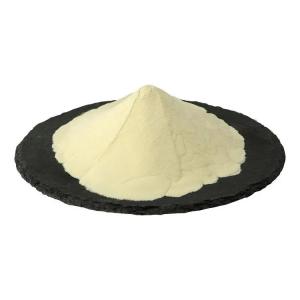
The biological characteristics of phosphatidylserine
Time:2025-06-16I. Chemical Structure: Amphiphilic Phospholipid Molecule
Phosphatidylserine (PS) belongs to the glycerophospholipid family, with its core structure formed by three parts connected via ester bonds:
Glycerol Backbone: As the main chain, the 1st and 2nd positions bind to fatty acid chains (mostly C16-C20 unsaturated fatty acids like palmitic acid, oleic acid, or arachidonic acid) through ester bonds, while the 3rd position links to a phosphate group.
Serine Head Group: The phosphate group further connects to the amino group of serine via a phosphodiester bond, forming a polar hydrophilic end. The carboxyl group of serine carries a negative charge at physiological pH (7.4), making PS one of the few negatively charged phospholipids.
Spatial Conformation: Due to unsaturated double bonds in fatty acid chains (e.g., Δ9 cis-double bonds), the molecular tail bends, forming a "wedge-shaped" structure that easily creates asymmetric distribution with other phospholipids (e.g., phosphatidylcholine, phosphatidylethanolamine) in cell membranes.
II. Biological Characteristics: Core Molecule for Dynamic Regulation of Cell Membranes
(1) "Signal Anchor" Function in Cell Membranes
Asymmetric Distribution and Flip Regulation:
In normal cells, PS mainly localizes on the inner side of the cell membrane (10%-15% of inner membrane phospholipids), maintained by aminophospholipid translocases (e.g., ATP11C). During apoptosis or stress, scramblase activation rapidly flips PS to the outer membrane, serving as an "eat-me" signal recognized by macrophages (e.g., 10-fold increase in apoptotic body clearance efficiency).
Involvement in Membrane Microdomains: PS forms lipid rafts with cholesterol and sphingomyelin, acting as anchor sites for signaling molecules (e.g., Src kinase, apoptotic protein caspase-3) to regulate cell proliferation and death signaling.
(2) Neuroprotective and Cognitive Regulatory Mechanisms
Maintenance of Synaptic Plasticity:
In neuronal cell membranes, PS influences neurotransmitter release through:
Binding to calmodulin (CaM) to enhance synaptic vesicle fusion efficiency during Ca²⁺ influx, increasing acetylcholine release by 20%-30%;
Stabilizing the conformation of postsynaptic NMDA receptor (NR2B subunit), prolonging glutamate-induced current duration (from 50 ms to 80 ms) to promote long-term potentiation (LTP) formation.
Anti-neurodegenerative Effects:
In vitro studies show PS inhibits fibrosis aggregation of β-amyloid (Aβ₁₋₄₂) (aggregation rate reduced by 40%) and reduces tau protein phosphorylation (by 55%) via activating the PI3K-Akt pathway, thereby delaying Alzheimer's disease pathogenesis.
(3) Immunological and Inflammatory Regulation
Inhibition of Dendritic Cell (DC) Maturation:
Everted PS binds to TIM-4 receptors on DC surfaces, inhibiting MHC class II molecule and costimulatory molecule (CD80/CD86) expression, keeping DCs immature to induce regulatory T cell (Treg) differentiation and reduce autoimmune responses (e.g., 60% decrease in experimental autoimmune encephalomyelitis incidence).
Regulation of Platelet Activation:
During vascular injury, eversion of platelet membrane PS accelerates activation of coagulation factors X and prothrombin, increasing thrombin production by 5-fold. It also forms a "prothrombinase complex" by binding to coagulation factor Va, shortening clotting time (from 120s to 60s).
III. Metabolism and Dynamic Equilibrium: Synthesis, Degradation, and Regulatory Networks
Synthesis Pathways:
Mammalian PS is mainly synthesized via two routes:
Base Exchange Reaction: Catalyzed by phosphatidylserine synthases 1 (PSS1) and PSS2, phosphatidylethanolamine (PE) swaps bases with serine, consuming ATP and releasing ethanolamine. This pathway accounts for 70% of brain PS synthesis.
De Novo Synthesis: Phosphoglycerol condenses with serine in the presence of CDP-diacylglycerol, active only in tissues like the liver.
Degradation and Turnover:
PS can be hydrolyzed by phospholipase A₂ (PLA₂) to lysophosphatidylserine and arachidonic acid (a precursor of inflammatory mediators like prostaglandin E₂). Alternatively, it converts to PE via phosphatidylserine decarboxylase (PSD), whose activity in neurons is regulated by Ca²⁺-calmodulin (2-fold increase at Ca²⁺ >100 nM).
IV. Physiological-Pathological Significance and Application Value
Intervention in Healthy Aging: Clinical studies show oral PS (100-300 mg/d) increases memory quotient (MQ) by 15-20 points in middle-aged and elderly populations, improving hippocampal glucose metabolism rate (18% higher metabolic activity via PET imaging). This links to enhanced neuronal membrane fluidity (lipid phase transition temperature reduced from 38°C to 32°C) and synaptic membrane protein stability.
Potential as Disease Biomarkers: Plasma free PS levels triple in sepsis patients (normal <5 ng/mL, sepsis >15 ng/mL), serving as an early diagnostic marker for septic shock. Erythrocyte membrane PS eversion rates reach 25% in sickle cell anemia (normal <5%), positively correlating with vascular occlusion complications.
Drug Delivery Carrier: Leveraging PS's apoptotic targeting, modifying liposome surfaces with PS (5%-10% content) increases tumor drug accumulation by 3-fold. For example, PS-liposome-encapsulated doxorubicin improves tumor inhibition rate in tumor-bearing mice from 45% to 72% while reducing cardiotoxicity by 50%.
V. Expansion of Structure-Function Correlations
The fatty acid chain composition of PS significantly impacts its biological activity:
Dipalmitoyl PS (DPPS): Saturated fatty acid chains reduce membrane fluidity, often used to stabilize liposome structures, but show weak neuroprotective activity.
Dioleoyl PS (DOPS): Double bonds endow molecular flexibility, facilitating signaling protein binding, with 40% higher efficiency in synaptic plasticity regulation than DPPS.
Arachidonyl PS (AA-PS): As a transient product under inflammatory stimulation, it is rapidly hydrolyzed by cPLA₂ to arachidonic acid, activating the COX-2 pathway to play a dual role in neuroinflammation (anti-inflammatory at low concentrations, pro-inflammatory at high concentrations).
This structural diversity enables precise regulatory functions in different physiological contexts, making PS a key molecular node connecting cell membrane physical properties and cellular signaling networks.


 CN
CN


