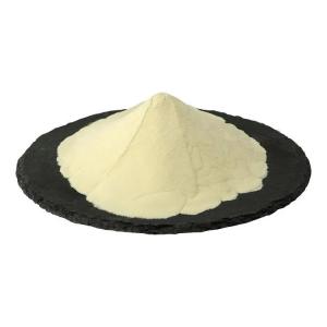
The distribution of phosphatidylserine in the human body
Time:2025-06-20I. Distribution Characteristics of Phosphatidylserine (PS) in the Human Body
As a glycerophospholipid containing serine, phosphatidylserine distributes unevenly in various human tissues, with its content closely related to tissue metabolic activity and cellular structural complexity:
Brain: Constitutes 15%–20% of total brain phospholipids, with extremely high concentrations in the cerebral cortex, hippocampus, synaptic vesicles, and other regions, serving as one of the unique phospholipid components in the central nervous system.
Peripheral Tissues: Lower content (1%–5% of total phospholipids) in liver, kidney, muscle, etc., mainly localized in the inner leaflet of cell membranes to participate in cell signal transduction and apoptosis regulation.
Blood and Immune Cells: Higher PS content on the cell membranes of platelets and lymphocytes plays a key role in coagulation and immune recognition (e.g., PS exposure on apoptotic cells acts as a "phagocytic signal").
II. Biological Mechanisms of High Phosphatidylserine Content in the Brain
The specific enrichment of brain phosphatidylserine directly relates to its unique physiological functions and structural requirements, analyzable from the following dimensions:
1. Structural Basis of Neural Cell Membranes
Material Support for Synaptic Plasticity: Brain neurons form complex networks via synaptic connections. The cell membranes of synaptic vesicles require high fluidity and flexibility to support synaptogenesis, neurotransmitter release, and dynamic receptor regulation. The negatively charged serine head and unsaturated fatty acid chains (e.g., arachidonic acid) in PS form "microdomains" in the membrane, maintaining the liquid-ordered phase of synaptic membranes to provide a structural basis for neural signal transmission.
Myelin and Axon Protection: Myelin in the central nervous system, formed by oligodendrocytes, wraps axons to accelerate nerve impulse conduction. PS enrichment in myelin enhances membrane insulation and damage resistance, while its negative charge binds to myelin proteins (e.g., myelin basic protein) to maintain myelin structural stability.
2. Core Role in Neural Signal Transduction
Calcium Signaling and Enzyme Activity Regulation: Processes such as excitation-contraction coupling and synaptic vesicle exocytosis in brain neurons rely on calcium (Ca²⁺) signals. As a negatively charged phospholipid in the inner membrane, PS binds to Ca²⁺ to form a "calcium-PS" complex, recruiting and activating signaling molecules like protein kinase C (PKC) and phospholipase C (PLC) to regulate neurotransmitter synthesis and release. For example, PS-mediated Ca²⁺ signals are crucial for long-term potentiation (LTP, the molecular basis of learning and memory) in hippocampal neurons.
Anchoring of Receptors and Ion Channels: Many neurotransmitter receptors (e.g., γ-aminobutyric acid receptor GABAR, NMDA receptor) and ion channels (e.g., voltage-gated calcium channels) require binding to PS to maintain correct conformation and function. PS embeds into receptor transmembrane domains via hydrophobic interactions or binds to intracellular receptor domains via electrostatic interactions, ensuring their localization and activity on cell membranes.
3. Essential Substrate for Neuron Development and Repair
Neurogenesis and Synaptogenesis: During embryonic development, brain PS content rises sharply with neuron proliferation and synaptic network formation. As a precursor, PS is hydrolyzed by phospholipases to produce serine and arachidonic acid, with the latter converted into prostaglandins and leukotrienes to participate in neural cell differentiation and synaptic connection establishment. Studies show mice lacking PS synthase exhibit abnormal cerebral cortical neuron migration and reduced synaptic numbers.
Damage Repair and Anti-Apoptotic Mechanisms: Since brain neurons are non-renewable, PS participates in neuroprotection through two pathways:
Membrane repair: When neurons are damaged by oxidative stress or inflammation, PS acts as a "membrane patch" to fill damaged areas and maintain membrane integrity.
Anti-apoptotic signaling: Normally located in the inner membrane, PS externalizes during apoptosis. However, high PS content in brain neurons competitively inhibits the membrane localization of apoptosis-related proteins (e.g., Bax), delaying the apoptotic process.
4. Blood-Brain Barrier and Metabolic Specificity
Active Transport and Local Synthesis: Due to its large molecular weight and charge, PS cannot passively diffuse through the blood-brain barrier. Brain PS is mainly obtained via:
Local synthesis: PS synthases (e.g., PSS1, PSS2) in astrocytes and neurons use serine and phosphatidylethanolamine (PE) as substrates to synthesize PS on mitochondria-associated membranes (MAM).
Transporter-mediated transport: Blood-brain barrier endothelial cells express PS transporters (e.g., ATP11C) to reverse-transport peripheral PS into the brain, though less efficiently than local synthesis.
Energy Adaptation to High Metabolic Demand: The brain, accounting for 2% of body weight, consumes 20% of systemic oxygen and glucose. Its high metabolic rate relies on efficient energy conversion. PS's unsaturated fatty acid chains serve as components of mitochondrial membranes to optimize oxidative phosphorylation efficiency, while its degradation products (e.g., serine) enter the one-carbon metabolic pathway to provide extra energy substrates for neurons.
III. Dysfunction and Disease Association
Reduced brain PS content links to multiple neurodegenerative diseases. For example, PS levels in the hippocampus of Alzheimer's disease (AD) patients decrease by 30%–40%, potentially causing:
Decreased synaptic membrane fluidity and neurotransmitter release disorders;
Accelerated aggregation of Aβ amyloid proteins (PS deficiency impairs Aβ clearance);
Excessive phosphorylation of Tau protein, promoting neurofibrillary tangle formation.
Clinical studies show exogenous PS supplementation (e.g., soybean-derived PS) improves cognitive function in AD model animals, further confirming the physiological significance of high brain PS content.
IV. Conclusion
The specific enrichment of phosphatidylserine in the brain represents an adaptive outcome of nervous system evolution, driven by its structural characteristics, signal regulatory functions, and neuroprotective effects. This distribution not only meets the brain's high-complexity structural needs but also provides a material basis for neuronal information processing, development, and repair, serving as an essential component of the central nervous system's precise regulatory mechanisms.


 CN
CN


