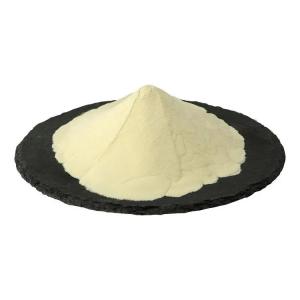
Phosphatidylserine is an important membrane phospholipid
Time:2025-06-18Phosphatidylserine (PS) is a vital membrane phospholipid that plays a pivotal role in cellular physiology. The following analysis explores its biological properties from chemical structure, biosynthesis, membrane distribution, and physiological functions:
I. Chemical Structure: Polar Molecular Characteristics of the Phospholipid Family
1. Basic Composition and Bonding Patterns
Skeletal Structure: Built around a glycerol core, ester-bonded to two fatty acid chains (R₁, R₂) and a phosphate group, which further links to the amino group of serine via a phosphoester bond, forming a "glycerol-fatty acid-phosphate-serine" architecture.
Fatty Acid Chain Features: Predominantly unsaturated fatty acids (e.g., palmitic acid, oleic acid, arachidonic acid) with 16–20 carbon atoms. Unsaturated double bonds endow molecular flexibility, influencing membrane fluidity.
Polar Head and Hydrophobic Tail: The serine-phosphate moiety forms a polar hydrophilic head, while fatty acid chains constitute a hydrophobic tail. This amphipathic structure makes it a fundamental building block of biological membranes.
2. Stereochemistry and Isomers
Natural PS features an L-configuration glycerol backbone (sn-1 and sn-2 positions bind fatty acids), with the sn-2 position often conjugated to long-chain polyunsaturated fatty acids (e.g., DHA), conferring unique biological activity.
II. Biosynthesis: A Dynamically Regulated Multifaceted Process
1. Major Synthetic Pathways
CDP-Diacylglycerol Pathway: In bacteria and plants, PS is synthesized from CDP-diacylglycerol (cytidine diphosphate diacylglycerol) and serine, catalyzed by PS synthase, with cytidine monophosphate (CMP) released.
Base Exchange Pathway: In mammals, primarily via base exchange between phosphatidylethanolamine (PE)/phosphatidylcholine (PC) and serine, mediated by PS synthases 1 (PSS1) and 2 (PSS2), dependent on mitochondrial-associated membrane (MAM) localization.
Salvage Pathway: Minor PS is synthesized by direct conjugation of serine to existing phospholipid groups or reacylation of lysophosphatidylserine.
2. Subcellular Localization and Regulation
Synthesis occurs mainly in the endoplasmic reticulum (ER) and MAM, with subsequent transport to other organelles (e.g., cell membrane, Golgi) via phospholipid transfer proteins (e.g., PITP) or membrane fusion.
Calcium ions (Ca²⁺) activate phosphatidylserine decarboxylase (PSD), promoting PS conversion to PE for dynamic membrane phospholipid regulation.
III. Membrane Distribution: Asymmetry and Physiological Significance
1. Asymmetric Localization in Cell Membranes
In normal cells, PS primarily resides in the inner leaflet (cytoplasmic side) of the plasma membrane, accounting for 10%–20% of inner leaflet phospholipids, with <5% in the outer leaflet. This distribution is regulated by:
Flippase (e.g., ATP11C): Consumes ATP to transport PS from the outer to inner leaflet.
Scramblase (e.g., TMEM16 family): Non-specifically flips phospholipids upon Ca²⁺ activation, disrupting asymmetry.
2. The "Signal Flag" of Apoptosis
During apoptosis, elevated Ca²⁺ activates scramblase and inhibits flippase, causing PS externalization to the outer membrane leaflet. Externalized PS is recognized by scavenger receptors (e.g., TIM4, Bai1) on phagocytes, mediating apoptotic cell clearance and preventing inflammation.
IV. Biological Properties and Physiological Functions: From Membrane Structure to Signal Transduction
1. Modulator of Membrane Structure and Fluidity
The negatively charged headgroup (ionized serine carboxyl) interacts with positively charged regions of membrane proteins (e.g., histidine, lysine residues), influencing protein conformation and activity (e.g., GPCRs, ion channels).
Unsaturated fatty acid chains endow membrane fluidity, while PS aggregation (via charge interactions) regulates membrane microdomain (e.g., lipid raft) formation for signal complex assembly.
2. Key Participant in Signal Transduction
Apoptotic Signaling: PS externalization is an early apoptotic event, recruiting apoptotic proteins (e.g., caspase-3) to the membrane for apoptotic body formation, in addition to serving as a phagocytic signal.
Coagulation Function: Activated platelets expose PS, providing a binding platform for coagulation factors (e.g., factor X, factor V), accelerating prothrombin conversion to thrombin for hemostasis.
Neuronal Signal Regulation: In neurons, PS enriches presynaptic membranes and synaptic vesicles, enhancing calmodulin (CaM)-protein kinase C (PKC) binding to modulate learning- and memory-related signaling.
3. Roles in Special Physiological Scenarios
Embryonic Development: PS is critical for early embryonic cell division and differentiation; knockout of PS synthase genes leads to embryonic lethality in mice.
Immune Regulation: PS on dendritic cell (DC) surfaces modulates T-cell activation, while macrophages secreting anti-inflammatory cytokines (e.g., IL-10) after phagocytosing PS-bearing apoptotic cells suppress excessive immune responses.
V. Metabolism and Degradation: Maintaining Membrane Homeostasis
PS is hydrolyzed by phospholipase A₂ (PLA₂) to lysophosphatidylserine and free fatty acids (e.g., arachidonic acid for inflammatory signaling).
It converts to PE via PSD or participates in mitochondrial phospholipid metabolism through the PSD pathway.
VI. Association with Diseases: Bridging Physiology and Pathology
Neurodegenerative Diseases: PS content significantly declines in the brains of Alzheimer’s disease (AD) patients. Exogenous PS supplementation (e.g., soybean- or bovine brain-derived PS) improves cognitive function, possibly by maintaining synaptic membrane stability.
Cardiovascular Diseases: Abnormal PS externalization promotes platelet over-activation and thrombosis, while impaired PS clearance from apoptotic cells may trigger autoimmune diseases (e.g., systemic lupus erythematosus).
The chemical structure of PS dictates its amphipathicity and charge properties, making it a structural basis and signaling hub for biological membranes. From membrane asymmetry to apoptotic signaling, neuronal regulation to disease pathogenesis, PS’s biological characteristics permeate multiple layers of cellular life activities. In-depth understanding of structure-function relationships not only provides an entry point for membrane biology research but also lays a theoretical foundation for preventing and treating related diseases (e.g., PS supplement applications).


 CN
CN


